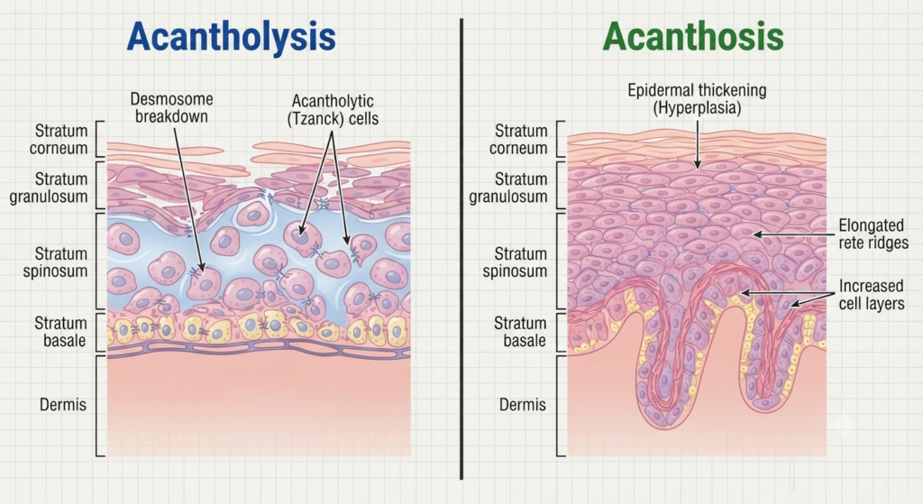
The main difference between Acantholysis vs. Acanthosis is that acantholysis refers to the loss of adhesion between epidermal skin cells (keratinocytes), causing them to separate and often resulting in blisters or erosions, whereas acanthosis refers to the thickening of the epidermis, particularly the stratum spinosum, due to increased proliferation of keratinocytes. Acantholysis is commonly associated with autoimmune blistering disorders such as pemphigus vulgaris, while acanthosis is frequently linked to insulin resistance, obesity, hormonal disorders, and chronic skin irritation.
Although both conditions affect the epidermis and are important findings in dermatology, they differ significantly in their causes, microscopic appearance, symptoms, diagnosis, treatment, and clinical significance. Understanding Acantholysis vs. Acanthosis helps medical students, healthcare professionals, and patients better recognize various skin disorders and their underlying mechanisms.
Acantholysis vs. Acanthosis Comparison Table
The table below summarizes the major differences between Acantholysis vs. Acanthosis.
| Feature | Acantholysis | Acanthosis |
|---|---|---|
| Definition | Loss of adhesion between epidermal keratinocytes | Thickening of the epidermis due to increased keratinocyte proliferation |
| Primary Change | Separation of skin cells | Increased thickness of the epidermis |
| Epidermal Layer Affected | Mainly the spinous layer with cell separation | Primarily the stratum spinosum |
| Mechanism | Breakdown of desmosomes | Hyperplasia of epidermal cells |
| Main Cause | Autoimmune diseases, infections, genetic disorders | Insulin resistance, obesity, hormonal disorders, friction |
| Skin Appearance | Blisters, erosions, ulcers | Thickened, velvety, rough skin |
| Cell Adhesion | Lost | Preserved |
| Histopathology | Acantholytic (detached) keratinocytes | Thickened epidermis with elongated rete ridges |
| Common Diseases | Pemphigus vulgaris, Pemphigus foliaceus | Acanthosis nigricans, chronic eczema |
| Symptoms | Painful blisters and skin erosion | Usually painless skin thickening |
| Diagnosis | Skin biopsy, immunofluorescence | Clinical examination and biopsy if required |
| Treatment | Immunosuppressive therapy | Treat underlying metabolic or hormonal disorder |
What Is Acantholysis?
Acantholysis is a pathological condition characterized by the loss of cohesion between keratinocytes, the primary cells of the epidermis. Normally, these cells are tightly connected by specialized structures called desmosomes, which maintain the strength and integrity of the skin. When these cell-to-cell connections are disrupted, the keratinocytes separate from one another, creating spaces within the epidermis that eventually develop into blisters, vesicles, or erosions.
Acantholysis is not a disease itself but rather a microscopic pathological finding observed in several dermatological disorders. It is most commonly associated with autoimmune blistering diseases, especially pemphigus vulgaris, in which autoantibodies attack desmosomal proteins responsible for holding epidermal cells together.
The severity of acantholysis depends on the underlying cause and may range from localized skin involvement to widespread blistering affecting both the skin and mucous membranes.
Characteristics of Acantholysis
- Loss of adhesion between keratinocytes
- Breakdown of desmosomes
- Formation of intraepidermal clefts
- Development of fragile blisters
- Commonly associated with autoimmune diseases
- May involve skin and oral mucosa
- Produces acantholytic cells under microscopy
- Causes painful erosions after blister rupture
- Usually requires prompt medical treatment
- Can become life-threatening if untreated
Common Causes of Acantholysis
- Pemphigus vulgaris
- Pemphigus foliaceus
- Hailey-Hailey disease
- Darier disease
- Viral infections (occasionally)
- Certain drug-induced skin reactions
What Is Acanthosis?
Acanthosis refers to the abnormal thickening of the epidermis, particularly the stratum spinosum, caused by an increased number of keratinocytes. Unlike acantholysis, there is no separation of skin cells. Instead, the epidermis becomes thicker due to excessive cell proliferation.
The most well-known clinical condition associated with this process is acanthosis nigricans, which presents as dark, velvety, thickened skin, commonly affecting the neck, armpits, groin, elbows, and other body folds. Acanthosis is often considered a marker of underlying metabolic disorders such as insulin resistance, obesity, type 2 diabetes, and hormonal imbalances.
Although acanthosis itself is usually harmless, it may indicate an underlying medical condition that requires evaluation and treatment.
Characteristics of Acanthosis
- Thickening of the epidermis
- Increased proliferation of keratinocytes
- Primarily affects the stratum spinosum
- Cell-to-cell adhesion remains intact
- Skin appears velvety or rough
- Often associated with hyperpigmentation
- Usually develops gradually
- Commonly linked to insulin resistance
- Frequently occurs in body folds
- Generally non-blistering and painless
Common Causes of Acanthosis
- Obesity
- Insulin resistance
- Type 2 diabetes
- Polycystic ovary syndrome (PCOS)
- Hormonal disorders
- Chronic friction
- Certain medications
- Rarely, internal malignancies
Acantholysis vs. Acanthosis: Key Differences
1. Definition
The most fundamental difference between Acantholysis vs. Acanthosis lies in the type of change that occurs within the epidermis. Although both conditions affect the outer layer of the skin, they involve entirely different pathological processes and lead to different clinical manifestations.
Acantholysis is the loss of adhesion between keratinocytes, the primary cells of the epidermis. This occurs when the specialized intercellular junctions known as desmosomes become damaged or destroyed, causing neighboring skin cells to separate from one another. As the cells lose contact, spaces develop within the epidermis, eventually forming fragile blisters or erosions. Acantholysis is therefore considered a cell separation disorder and is commonly seen in autoimmune blistering diseases.
In contrast, Acanthosis is the abnormal thickening of the epidermis, particularly the stratum spinosum (prickle cell layer). Instead of losing adhesion, the keratinocytes multiply more rapidly than normal, increasing the thickness of the skin. The cells remain firmly attached, and blister formation does not occur. Clinically, acanthosis usually presents as thickened, rough, or velvety skin rather than fragile blisters.
In simple terms, acantholysis involves the separation of epidermal cells, whereas acanthosis involves an increase in the number of epidermal cells.
2. Underlying Mechanism
The biological mechanism responsible for these two conditions is entirely different, which explains their distinct clinical appearances.
In acantholysis, the primary defect involves the destruction or dysfunction of desmosomes, which are protein-rich structures responsible for holding keratinocytes together. In autoimmune diseases such as pemphigus vulgaris, antibodies attack desmosomal proteins (especially desmogleins), causing keratinocytes to lose their adhesion. As a result, the cells detach from one another, creating intraepidermal clefts that later develop into fluid-filled blisters.
In acanthosis, there is no damage to cell junctions. Instead, the epidermis becomes thicker because keratinocytes undergo excessive proliferation. Factors such as elevated insulin levels, hormonal disturbances, chronic irritation, or inflammatory stimuli stimulate epidermal growth, producing a thicker stratum spinosum. Since the intercellular junctions remain intact, the skin maintains its structural integrity despite becoming thicker.
Thus, the underlying mechanism in acantholysis is loss of cell adhesion, whereas the mechanism in acanthosis is increased cell growth and proliferation.
3. Effect on Skin Structure
The impact of these conditions on the architecture of the skin differs considerably.
Acantholysis weakens the structural integrity of the epidermis because the keratinocytes no longer adhere to each other. The separated cells create empty spaces within the epidermis, making the skin extremely fragile. Even minor friction or trauma can rupture the resulting blisters, leading to painful erosions, ulcers, crust formation, and an increased risk of secondary bacterial infection. In severe autoimmune diseases, extensive skin loss may even resemble burn injuries.
Acanthosis, on the other hand, increases the thickness of the epidermis without compromising cell adhesion. Instead of becoming fragile, the affected skin appears thicker, rougher, and often velvety. Although acanthosis may alter the skin’s appearance and texture, it generally does not reduce its mechanical strength or cause blister formation.
Therefore, while acantholysis causes structural weakening of the skin, acanthosis results in epidermal thickening with preserved structural integrity.
4. Epidermal Changes
Microscopic examination reveals strikingly different histological features in these two conditions.
In acantholysis, histopathological examination shows detached, rounded keratinocytes suspended within intraepidermal spaces. These detached cells are known as acantholytic cells and represent one of the hallmark findings of autoimmune blistering disorders. Depending on the disease, the separation may occur above the basal layer or within other levels of the epidermis.
In acanthosis, microscopic examination demonstrates hyperplasia of the stratum spinosum, resulting in a noticeably thickened epidermis. The rete ridges become elongated and broadened, while the keratinocytes remain firmly attached through intact desmosomes. No intraepidermal clefts or detached keratinocytes are observed.
Consequently, acantholysis is identified by cell separation, whereas acanthosis is recognized by epidermal hyperplasia and increased epidermal thickness.
5. Causes
The underlying causes of acantholysis and acanthosis are very different because each condition results from a distinct pathological process.
Acantholysis most commonly develops due to autoimmune disorders, where the body’s immune system mistakenly attacks proteins responsible for maintaining cell-to-cell adhesion. It may also occur in inherited disorders affecting desmosomal proteins, certain viral infections, severe inflammatory skin diseases, and adverse drug reactions. Autoimmune blistering disorders remain the most frequent cause of clinically significant acantholysis.
Acanthosis, in contrast, is primarily associated with metabolic and endocrine abnormalities. Conditions such as obesity, insulin resistance, type 2 diabetes mellitus, polycystic ovary syndrome (PCOS), growth hormone disorders, thyroid dysfunction, and prolonged skin friction commonly stimulate excessive epidermal growth. Rarely, sudden onset of widespread acanthosis may serve as a paraneoplastic sign associated with internal malignancies, particularly gastrointestinal cancers.
Thus, acantholysis is mainly linked to immune-mediated skin disorders, whereas acanthosis is commonly associated with metabolic, hormonal, and systemic conditions.
6. Clinical Appearance
The physical appearance of these conditions differs so markedly that clinicians can often suspect the diagnosis during the initial examination.
Acantholysis typically presents with thin-walled, fragile blisters that rupture easily, leaving painful erosions and crusted lesions. The affected skin appears raw and moist after blister rupture. In autoimmune diseases such as pemphigus vulgaris, lesions frequently involve both the skin and mucous membranes, including the mouth, throat, and nose. Gentle rubbing of apparently normal skin may even induce blister formation, a phenomenon known as Nikolsky’s sign.
Acanthosis presents as darkened, thickened, velvety plaques that develop gradually over months or years. These lesions most commonly appear on the neck, armpits, groin, elbows, knees, knuckles, and other skin folds. Unlike acantholysis, the skin surface remains intact without blistering or ulceration.
Therefore, acantholysis produces fragile blistering skin, while acanthosis produces thickened, hyperpigmented skin without blisters.
7. Symptoms
The symptoms experienced by patients differ according to the underlying pathological process.
Patients with acantholysis often experience pain, burning, tenderness, and significant discomfort because the protective epidermal barrier is disrupted. Oral lesions may interfere with eating, drinking, and speaking. Extensive blistering can lead to fluid loss, electrolyte imbalance, infection, and reduced quality of life if not treated promptly.
In contrast, acanthosis is usually asymptomatic. Most patients seek medical attention because of cosmetic concerns related to skin darkening or thickening. Mild itching or irritation may occur in some individuals, particularly where friction is present, but pain is uncommon.
Overall, acantholysis tends to produce painful and potentially serious symptoms, whereas acanthosis generally causes minimal physical discomfort.
8. Diseases Associated
Both conditions serve as important pathological findings in dermatology, but they are associated with different groups of diseases.
Acantholysis is a characteristic feature of several blistering disorders, including:
- Pemphigus vulgaris
- Pemphigus foliaceus
- Hailey-Hailey disease
- Darier disease
- Grover’s disease (Transient acantholytic dermatosis)
- Some viral skin infections
- Drug-induced blistering disorders
Acanthosis commonly occurs in association with:
- Acanthosis nigricans
- Obesity
- Type 2 diabetes mellitus
- Insulin resistance syndrome
- Polycystic ovary syndrome (PCOS)
- Cushing syndrome
- Acromegaly
- Hypothyroidism
- Chronic eczema
- Internal malignancies (rare malignant acanthosis nigricans)
These associated diseases often provide important diagnostic clues regarding the underlying cause of the skin changes.
9. Diagnosis
Because these conditions have different causes and microscopic features, their diagnostic approaches also differ.
Diagnosis of acantholysis usually requires skin biopsy followed by histopathological examination to identify acantholytic keratinocytes and intraepidermal clefts. Direct immunofluorescence is considered the gold standard for diagnosing autoimmune blistering disorders because it detects antibodies deposited within the skin. Blood tests for circulating autoantibodies may also help confirm diseases such as pemphigus vulgaris.
Diagnosis of acanthosis often begins with a careful clinical examination, as the characteristic velvety thickening is usually recognizable. Laboratory investigations may include fasting blood glucose, HbA1c, fasting insulin levels, thyroid function tests, hormonal evaluation, or imaging studies if an underlying endocrine disorder or malignancy is suspected. Skin biopsy is reserved for atypical cases or when confirmation is required.
Thus, diagnosis of acantholysis focuses primarily on identifying autoimmune skin damage, whereas diagnosis of acanthosis emphasizes detecting underlying metabolic or endocrine disorders.
10. Treatment
Since the underlying causes differ completely, treatment strategies are also fundamentally different.
Acantholysis requires treatment of the disease responsible for destroying epidermal cell adhesion. Autoimmune blistering disorders are commonly managed with systemic corticosteroids, immunosuppressive drugs, biologic agents such as rituximab, topical therapies, wound care, pain management, and infection prevention. Early diagnosis and prompt treatment are essential because untreated autoimmune acantholysis can become life-threatening.
Acanthosis, by contrast, is managed by correcting the underlying metabolic or hormonal abnormality. Weight reduction, improved insulin sensitivity, blood glucose control, treatment of endocrine disorders, and discontinuation of causative medications often lead to gradual improvement. Topical retinoids, keratolytic agents, chemical peels, and laser therapy may also be used to improve skin appearance when necessary.
In summary, acantholysis treatment targets the underlying disease causing cell separation, whereas acanthosis treatment focuses on correcting the metabolic or hormonal factors responsible for epidermal thickening.
Similarities Between Acantholysis and Acanthosis
Although Acantholysis and Acanthosis represent different pathological processes, they share several important characteristics. Both conditions affect the epidermis, are commonly evaluated by dermatologists and pathologists, and may indicate underlying systemic diseases. Understanding their similarities helps clinicians differentiate them while recognizing their importance in diagnosing skin disorders.
Some key similarities include:
- Both are pathological changes involving the epidermis.
- Both are identified through clinical examination and histopathological analysis.
- Both affect keratinocytes, the primary cells of the epidermis.
- Both may occur as manifestations of underlying systemic diseases.
- Both can be associated with chronic dermatological disorders.
- Both may require skin biopsy for confirmation in uncertain cases.
- Both influence the appearance and structure of the skin.
- Both require treatment of the underlying cause rather than only treating skin changes.
- Both are important diagnostic findings in dermatopathology.
- Early diagnosis improves treatment outcomes and prevents complications.
Advantages of Understanding Acantholysis
Recognizing acantholysis is important because it serves as a key diagnostic clue for several potentially serious skin disorders. Early identification allows prompt treatment, reducing complications and improving patient outcomes.
Some important advantages of understanding acantholysis include:
- Helps diagnose autoimmune blistering diseases at an early stage.
- Enables timely initiation of immunosuppressive therapy.
- Prevents progression to extensive skin damage.
- Reduces the risk of secondary bacterial infections.
- Assists pathologists in interpreting skin biopsy findings.
- Improves differentiation between various blistering disorders.
- Helps monitor disease activity and treatment response.
- Supports appropriate wound care management.
- Improves patient prognosis through early intervention.
- Provides valuable information for dermatologists and medical students.
Advantages of Understanding Acanthosis
Understanding acanthosis is equally valuable because it often serves as an external marker of underlying metabolic or endocrine disorders. Recognizing these skin changes can lead to early detection of diseases that may otherwise remain undiagnosed.
Some advantages of understanding acanthosis include:
- Helps identify insulin resistance at an early stage.
- Supports early diagnosis of type 2 diabetes mellitus.
- Assists in detecting obesity-related skin changes.
- May indicate hormonal disorders such as PCOS.
- Encourages timely lifestyle modification.
- Helps monitor metabolic health.
- Improves cosmetic management of skin thickening.
- Provides clues to rare internal malignancies in sudden-onset cases.
- Guides physicians toward appropriate laboratory investigations.
- Promotes preventive healthcare through early screening.
Which Is More Clinically Significant—Acantholysis or Acanthosis?
Neither acantholysis nor acanthosis is inherently more important because each represents a different pathological process with distinct clinical implications. Their significance depends on the underlying condition responsible for the skin changes.
Acantholysis is generally considered more urgent because it is frequently associated with autoimmune blistering diseases such as pemphigus vulgaris, which can become life-threatening if left untreated. Extensive blistering may lead to severe pain, dehydration, electrolyte imbalance, infections, and significant damage to the skin barrier. Early diagnosis and prompt immunosuppressive treatment are therefore essential.
Acanthosis, on the other hand, is usually not life-threatening by itself. However, it often serves as an important warning sign of systemic conditions such as insulin resistance, obesity, type 2 diabetes, polycystic ovary syndrome (PCOS), or endocrine disorders. Identifying acanthosis can lead to earlier diagnosis and management of these underlying diseases, reducing long-term health complications.
In summary:
Choose acantholysis as the more clinically significant finding when:
- Evaluating blistering skin diseases.
- Diagnosing autoimmune disorders.
- Assessing painful erosive skin lesions.
- Preventing serious skin infections and fluid loss.
- Managing dermatological emergencies.
Choose acanthosis as the more significant finding when:
- Screening for diabetes or insulin resistance.
- Evaluating obesity-related skin changes.
- Investigating endocrine disorders.
- Detecting metabolic syndrome.
- Identifying possible paraneoplastic syndromes in rare cases.
Rather than considering one superior to the other, clinicians interpret each finding within the patient’s overall clinical picture to establish the correct diagnosis and treatment plan.
Conclusion
The comparison of Acantholysis vs. Acanthosis highlights two entirely different pathological changes that occur within the epidermis. Acantholysis involves the loss of adhesion between keratinocytes, resulting in fragile blisters, erosions, and disruption of the skin barrier. It is most commonly associated with autoimmune blistering disorders such as pemphigus vulgaris and requires prompt medical treatment to prevent serious complications.
In contrast, Acanthosis is characterized by thickening of the epidermis, particularly the stratum spinosum, due to increased keratinocyte proliferation. It usually presents as dark, velvety, thickened skin and is frequently linked to insulin resistance, obesity, diabetes mellitus, hormonal disorders, and chronic friction. Although acanthosis is generally benign, it often serves as an important clinical marker of underlying metabolic disease.
Understanding the differences between Acantholysis vs. Acanthosis enables healthcare professionals, students, and patients to recognize these skin changes, understand their causes, and seek appropriate evaluation and treatment. Early diagnosis not only improves dermatological care but may also help identify serious systemic diseases before complications develop.
Frequently Asked Questions (FAQs)
The primary difference between Acantholysis vs. Acanthosis is that acantholysis involves the loss of adhesion between keratinocytes, leading to blister formation, whereas acanthosis involves thickening of the epidermis due to increased keratinocyte proliferation without causing blisters.
Acantholysis itself is not a disease, but a pathological finding. It commonly occurs in autoimmune disorders such as pemphigus vulgaris and pemphigus foliaceus, where antibodies attack proteins responsible for cell-to-cell adhesion in the epidermis.
Acanthosis is most commonly caused by insulin resistance, obesity, type 2 diabetes, hormonal disorders such as polycystic ovary syndrome (PCOS), chronic friction, certain medications, and, in rare cases, internal malignancies.
Yes. In many cases, acanthosis improves when the underlying cause is treated. Weight loss, improved blood sugar control, treatment of hormonal disorders, and management of insulin resistance often reduce skin thickening and discoloration over time.
Diagnosis usually involves a skin biopsy, histopathological examination, and direct immunofluorescence, which helps detect autoimmune antibodies affecting desmosomes. Blood tests for specific autoantibodies may also assist in confirming the diagnosis.
Reference:
1. “Acanthosis.” An Overview | ScienceDirect Topics
Read Next: