The main difference between Lacunae vs. Lamellae is their structure and function within biological tissues. Lacunae are small spaces or cavities within a tissue matrix that typically contain specialized cells, such as osteocytes in bone or chondrocytes in cartilage, whereas lamellae are thin layers or plates of tissue or extracellular matrix arranged in a specific pattern. Thus, lacunae are primarily associated with cell housing, while lamellae are primarily associated with layered tissue organization and structural support.
Understanding Lacunae vs. Lamellae is important in anatomy, histology, biology, and medical studies because these terms describe different structural components of tissues. In bone, for example, lacunae contain osteocytes, while lamellae form organized layers of mineralized bone matrix that contribute to strength and resistance to mechanical stress.
Lacunae vs. Lamellae Comparison Table
The table below highlights the major differences between Lacunae vs. Lamellae.
| Feature | Lacunae | Lamellae |
|---|---|---|
| Definition | Small spaces or cavities within a tissue matrix | Thin layers or plates of tissue or extracellular matrix |
| Singular Form | Lacuna | Lamella |
| Primary Structure | A cavity or space | A thin layer or sheet |
| Common Location | Bone and cartilage | Bone and other biological structures |
| Bone Function | Houses osteocytes | Forms organized layers of bone matrix |
| Cells Present | Often contains specialized cells | May contain or surround cells depending on the tissue |
| Bone Arrangement | Located between or within lamellae | Surrounds the central canal in osteons or occurs as other bone layers |
| Shape | Usually small, flattened, or irregular cavity | Thin, layered, curved, or plate-like structure |
| Main Role | Provides a space for tissue cells | Provides structural organization and strength |
| Connected Structures | Connected by canaliculi in bone | Organized into layers around osteons or other bone structures |
| Typical Example | Osteocyte lacuna | Concentric bone lamella |
| Primary Concept | Cell-containing space | Layered tissue structure |
What Are Lacunae?
Lacunae are small spaces, cavities, or depressions present within the extracellular matrix of certain tissues. The term “lacuna” means a small cavity or empty space.
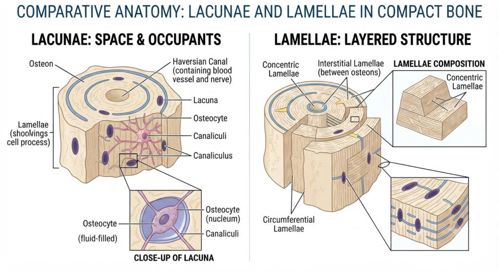
In bone tissue, lacunae are small spaces within the mineralized bone matrix that contain osteocytes, the mature cells of bone. Each osteocyte occupies a lacuna and extends cellular processes through tiny channels called canaliculi.
Lacunae are also found in cartilage. In cartilage, they contain chondrocytes, which are mature cartilage cells responsible for maintaining the cartilage matrix.
The structure of a lacuna therefore depends on the tissue in which it occurs. In bone, lacunae provide protected spaces for osteocytes, while in cartilage they provide spaces for chondrocytes.
Characteristics of Lacunae
Some important characteristics of lacunae include:
- Are small cavities or spaces within a tissue matrix.
- Commonly occur in bone and cartilage.
- House specialized cells.
- Bone lacunae contain osteocytes.
- Cartilage lacunae contain chondrocytes.
- Are surrounded by extracellular matrix.
- Bone lacunae are connected through canaliculi.
- Help maintain the health of the surrounding tissue.
- Are microscopic structures.
- Are important features in histological identification of bone and cartilage.
Examples of Lacunae
Examples of lacunae include:
- Osteocyte lacunae in bone.
- Chondrocyte lacunae in cartilage.
- Spaces containing mature bone cells.
- Spaces containing mature cartilage cells.
- Lacunae observed in microscopic sections of connective tissues.
What Are Lamellae?
Lamellae are thin layers, sheets, or plates of tissue or extracellular matrix. The term “lamella” refers to a thin layer or plate-like structure.
Lamellae are particularly important in bone tissue. In compact bone, lamellae are organized layers of mineralized bone matrix. In an osteon, concentric lamellae are arranged around a central canal containing blood vessels and nerves.
The collagen fibers in adjacent bone lamellae are arranged in different orientations. This organization contributes to the ability of bone to withstand mechanical forces from different directions.
Lamellae can also describe thin structures in other biological systems. For example, the gills of fish contain delicate lamellae that increase the surface area available for gas exchange.
Characteristics of Lamellae
Important characteristics of lamellae include:
- Are thin layers or plates.
- Can occur in bone and other biological structures.
- May contain extracellular matrix.
- In compact bone, they contain mineralized matrix.
- Form concentric layers within osteons.
- Contribute to the strength and organization of bone.
- Can increase surface area in certain biological structures.
- May have different arrangements depending on the tissue.
- Are important structural components of some organs.
- Can be observed microscopically.
Examples of Lamellae
Examples of lamellae include:
- Concentric lamellae in compact bone.
- Circumferential lamellae around the outer surface of bone.
- Interstitial lamellae between osteons.
- Gill lamellae in fish.
- Lamellar structures in various tissues and biological systems.
Lacunae vs. Lamellae: Key Differences
1. Basic Structure
The most important difference between Lacunae vs. Lamellae is their basic structural form.
A lacuna is a small cavity or space within a tissue matrix.
A lamella is a thin layer or plate of tissue or extracellular matrix.
Therefore:
Lacuna → Space
Lamella → Layer
This is the simplest structural distinction between the two terms.
2. Main Function
Lacunae and lamellae also have different primary functions.
Lacunae provide spaces for specialized cells. In bone, osteocytes occupy lacunae, while chondrocytes occupy lacunae in cartilage.
Lamellae provide organized layers of tissue. In bone, they contribute to structural strength and help distribute mechanical forces.
Thus, lacunae are mainly associated with cell location, while lamellae are mainly associated with tissue organization.
3. Location in Bone
Both structures are found in bone, but they occupy different positions.
Lacunae are small spaces within the bone matrix that house osteocytes.
Lamellae are layers of bone matrix that surround the central canal in an osteon or occur in other organized layers of bone.
In a typical osteon:
Central canal → Concentric lamellae → Lacunae containing osteocytes
This arrangement illustrates how the two structures work together within compact bone.
4. Relationship With Cells
Lacunae have a direct relationship with specialized cells.
An osteocyte resides inside a bone lacuna, while a chondrocyte resides inside a cartilage lacuna.
Lamellae, in contrast, are layers of tissue matrix. They are not primarily defined by the presence of a single cell.
Therefore:
Lacunae → House cells
Lamellae → Form tissue layers
5. Shape
The shapes of lacunae and lamellae are also different.
Lacunae are generally small, flattened, oval, or irregular spaces within the matrix.
Lamellae are thin layers, sheets, or plates. In compact bone, they can form curved concentric rings around a central canal.
Thus, lacunae have a cavity-like form, whereas lamellae have a layer-like form.
6. Role in Bone Strength
Lamellae have a major structural role in providing mechanical strength to bone.
The mineralized matrix and organized collagen fibers within bone lamellae help bone resist compression, tension, bending, and twisting.
Lacunae do not primarily provide mechanical strength. Their main function is to provide spaces for osteocytes, which maintain the surrounding bone matrix.
Therefore, lamellae are more directly associated with the structural organization of bone.
7. Connection With Canaliculi
Bone lacunae are connected with one another through microscopic channels called canaliculi.
These channels allow osteocytes to communicate and exchange nutrients and waste products through their cellular processes.
Lamellae, however, are not defined by canaliculi. Instead, they represent organized layers of bone matrix.
This makes canaliculi particularly relevant when studying lacunae.
8. Role in Osteons
Both lacunae and lamellae are important components of an osteon, but they perform different roles.
Lamellae form concentric layers around the central canal.
Lacunae are located between the layers of bone matrix and contain osteocytes.
Therefore, an osteon can be visualized as a highly organized structure containing:
Central canal → Lamellae → Lacunae → Canaliculi
This organization allows bone to combine structural strength with cellular maintenance.
9. Presence in Cartilage
Lacunae are a well-known feature of cartilage.
Chondrocytes are located within lacunae in the cartilage matrix. These spaces provide an environment in which the cells can maintain the surrounding extracellular matrix.
Lamellae are not a defining structural feature of ordinary cartilage in the same way that lacunae are.
Therefore, when studying cartilage histology, lacunae are particularly important for identifying chondrocytes.
10. Biological Significance
Lacunae are important for maintaining the cells embedded within specialized connective tissues.
Lamellae are important for creating organized tissue architecture and providing mechanical or functional advantages.
In bone, the two structures work together: lacunae house osteocytes, while lamellae organize the mineralized matrix around them.
Similarities Between Lacunae and Lamellae
Although Lacunae vs. Lamellae describes two different structures, they share several important characteristics.
Some major similarities include:
- Both are microscopic anatomical structures.
- Both can be observed in bone tissue.
- Both are important in histology.
- Both contribute to the organization of biological tissues.
- Both are associated with the extracellular matrix.
- Both help maintain the functional structure of bone.
- Both can be identified in microscopic sections.
- Both are important concepts in anatomy and biology.
- Both are associated with specialized tissue organization.
- Both can occur as components of complex biological structures.
Functions of Lacunae and Lamellae
The major functions of lacunae include:
- Providing spaces for specialized cells.
- Housing osteocytes in bone.
- Housing chondrocytes in cartilage.
- Protecting cells within the extracellular matrix.
- Supporting communication between osteocytes through canaliculi.
- Helping maintain bone and cartilage matrix.
- Providing a characteristic histological feature of bone and cartilage.
The major functions of lamellae include:
- Organizing the extracellular matrix into layers.
- Providing structural strength.
- Helping distribute mechanical stress.
- Supporting the architecture of compact bone.
- Forming concentric layers around osteonal central canals.
- Contributing to resistance against mechanical forces.
- Increasing functional surface area in some biological structures, such as gill lamellae.
Lacunae vs. Lamellae in Bone
Bone provides one of the clearest examples of the difference between lacunae and lamellae.
Compact bone is organized into structural units called osteons or Haversian systems. Each osteon contains a central canal surrounded by concentric layers of bone matrix called lamellae.
Osteocytes are located in small spaces called lacunae between the layers of mineralized matrix. The osteocytes extend processes through canaliculi, creating a communication network within the bone.
The basic arrangement can be represented as:
Central canal → Concentric lamellae → Lacunae → Osteocytes → Canaliculi
Lamellae provide structural organization and strength, while lacunae provide spaces for the cells that maintain the bone matrix.
This relationship is important for understanding the microscopic structure of compact bone.
Which Is More Important—Lacunae or Lamellae?
Neither lacunae nor lamellae is universally more important because they perform different functions.
Lacunae are essential for providing spaces for osteocytes and other specialized cells within certain connective tissues.
Lamellae are essential for organizing tissue matrix and providing structural strength, particularly in compact bone.
In bone, both structures work together. Osteocytes located in lacunae help maintain the surrounding bone matrix, while lamellae provide the organized structure that allows bone to withstand mechanical stress.
Therefore:
Choose lacunae when discussing cell-containing spaces in bone or cartilage.
Choose lamellae when discussing thin layers or organized sheets of tissue.
Conclusion
The comparison of Lacunae vs. Lamellae highlights two distinct structural features found in biological tissues. Lacunae are small spaces or cavities within a tissue matrix that house specialized cells such as osteocytes in bone and chondrocytes in cartilage. Lamellae, on the other hand, are thin layers or plates of tissue or extracellular matrix that provide organization and, in bone, contribute significantly to structural strength.
In compact bone, lacunae and lamellae work together as components of the osteon. Lamellae form organized layers around the central canal, while lacunae located within the matrix contain osteocytes. Canaliculi connect these lacunae and allow communication between osteocytes.
Understanding Lacunae vs. Lamellae is therefore important for studying bone histology, cartilage, anatomy, and connective tissue structure. The simplest distinction is that lacunae are spaces that house cells, whereas lamellae are layers that organize tissue structure.
Frequently Asked Questions (FAQs)
The primary difference between Lacunae vs. Lamellae is their structure. Lacunae are small spaces or cavities within a tissue matrix that commonly contain specialized cells, whereas lamellae are thin layers or plates of tissue or extracellular matrix.
A bone lacuna normally contains an osteocyte, which is a mature bone cell.
A cartilage lacuna normally contains a chondrocyte, which is a mature cartilage cell.
Lamellae are thin layers of mineralized bone matrix. In an osteon, concentric lamellae surround the central canal and contribute to the structural strength of compact bone.
Yes. Both lacunae and lamellae are important components of bone tissue. Lacunae contain osteocytes, while lamellae form organized layers of bone matrix.
References:
1. Birch, De B. “The Constitution and Relations of Bone Lamellæ, Lacunæ, and Canaliculi, and some Effects of Trypsin Digestion on Bone.” The Journal of Physiology, U.S. National Library of Medicine, July 1880
2. “Bone Tissue.” Bone Tissue – Structure and Functions of Human Tissue Types
3. Slomianka, Lutz. Blue Histology – Skeletal Tissues – Bone
Read Next:
- Difference Between Etiology and Pathophysiology
- Difference Between DNA Fingerprinting and DNA Profiling
- Difference Between Detritivores and Saprotrophs
- Difference Between Glenoid Cavity and Acetabulum
- Difference Between Mycoplasma and Mycobacterium
- Pinnatifid vs. Pinnatisect: Key Differences Explained
- Phosphodiester Bond vs. Phosphoester Bond
- Cloning vs. Subcloning
- Chemoorganotrophs vs. Chemolithotrophs
- Acantholysis vs. Acanthosis
- Flatbread vs. Pita